Subscribe to our newsletter for the latest news, events, and exclusive offers. Get updates straight to your inbox and be the first to know about promotions and discounts.
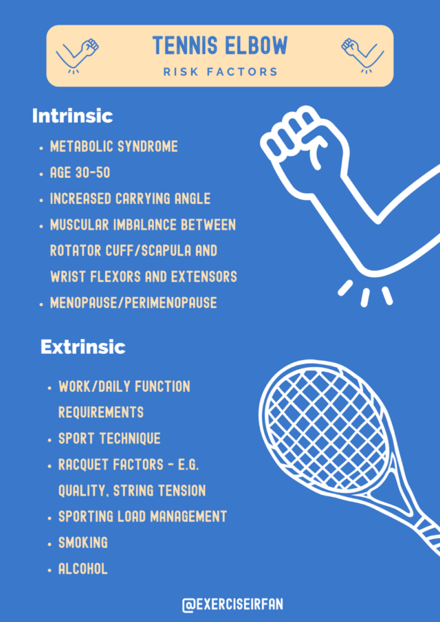
Tennis elbow (or lateral epicondylitis) is a common upper limb condition seen in the musculoskeletal (MSK) clinic. It can cause significant pain, restriction...
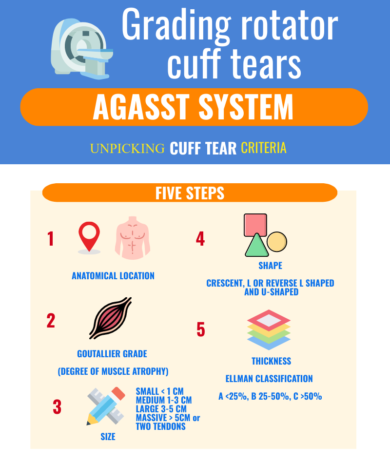
The rotator cuff muscles and tendons work as a dynamic structure to centralise the humeral head and aid movement...
Frozen shoulder (adhesive capsulitis) is a common condition within community musculoskeletal (MSK) interface services, which presents insidiously...
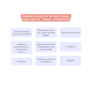
Ulnar nerve entrapment neuropathies commonly present to MSK and orthopaedics clinics, with patient’s...
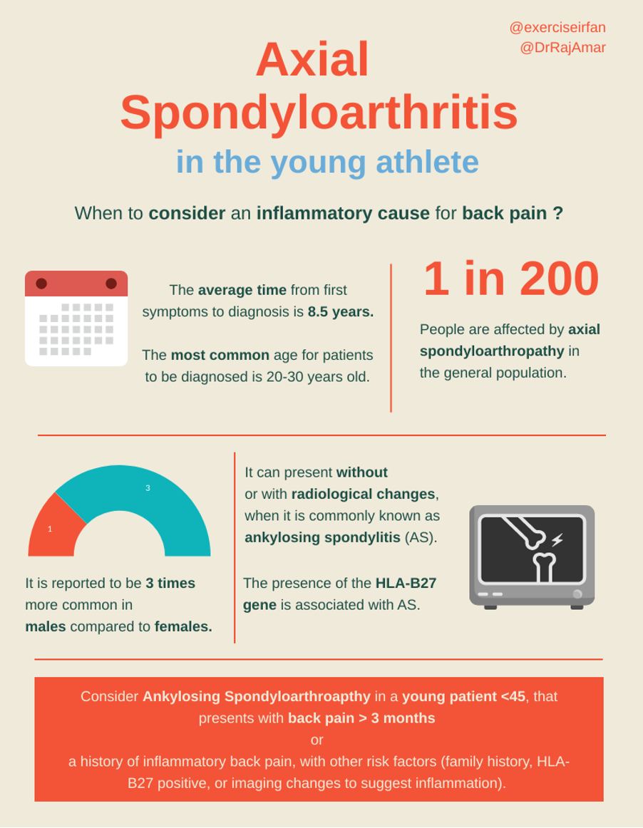
Back pain is a common complaint in the young adult athlete. This is...
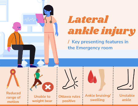
Ankle sprains affect anyone and everyone along the activity spectrum, from semi-professional players and athletes to weekend warriors and...
For the athlete or exercising patient, injury, training load, genetics, and training type have traditionally been...
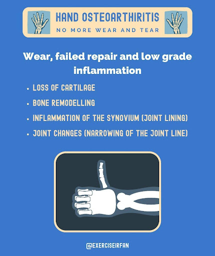
Hand osteoarthritis (OA) is a common presentation in the musculoskeletal (MSK) clinic, impacting millions of patients worldwide. It typically...
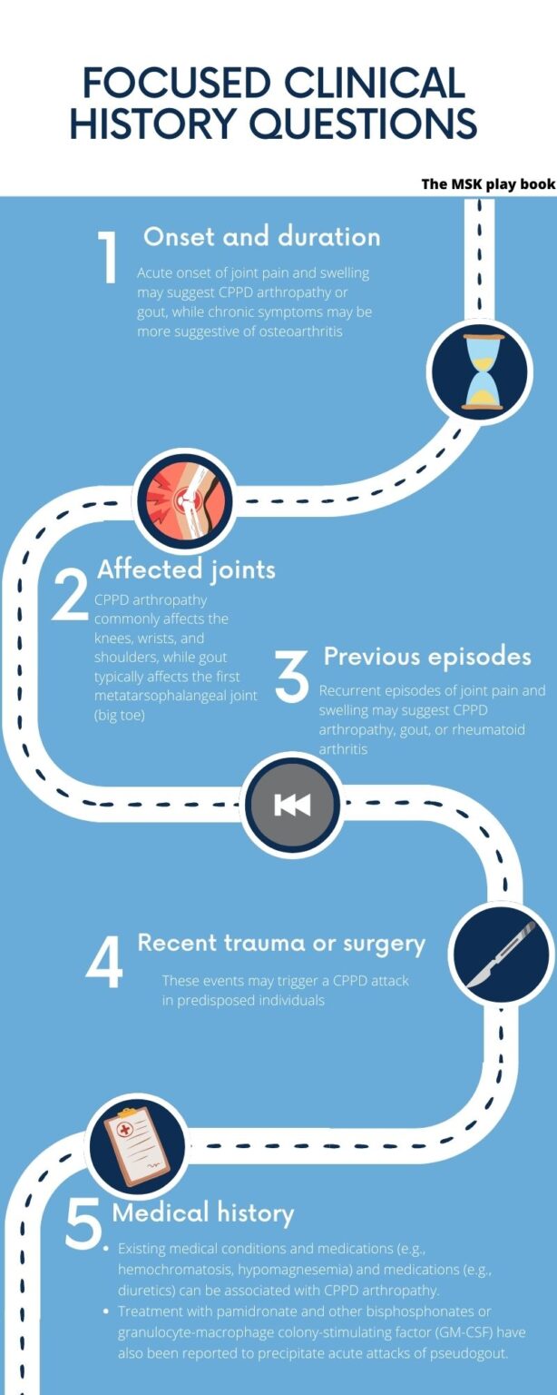
Chondrocalcinosis is a common imaging finding encountered in community MSK clinics. In some patients, it may be...
Bundle Includes Pre-Course Material:
Module 1: Learning to explain key MSK imaging report findings to a patient.
1.1 Understanding what shows up on each imaging sequence – your cheat codes for MRI/CT.
1.2 The MSK playbook, osteoarthritis, painful tendons, tears, and inflammatory conditions. What to look out for on each imaging modality.
1.3 - X-ray Quiz
Module 2: Common shoulder conditions in the MSK clinic
2.1 - The MSK playbook; Evidence updates on the frozen shoulder
2.2 – MRI of the shoulder – Live MRI tutorial to walk you through the key anatomy, how to identify common pathology and explain the scan to your patient!
2.3 – MSK imaging series - (Frozen Shoulder, Rotator Cuff Tendinopathy/Tears, Slap/Labral Injuries, Osteoarthritis, Dislocations)
2.4 MRI of the shoulder – Learn the key views to be able to show your patient
2.5 - The MSK playbook; Evidence update on rotator cuff tears
2.6 - The MSK playbook; Evidence update on rotator cuff tendinopathy
2.7 - The MSK playbook; Evidence update on calcific tendinopathy
Module 3: Common elbow conditions in the MSK clinic
3.1 The MSK playbook; Evidence update on common tendinopathies.
3.2 – MRI of the Elbow – Live MRI tutorial to walk you through the key anatomy, how to identify common pathology and explain the scan to your patient!
3.3 – MSK imaging series - (Tennis elbow, Golfers elbow, Cubital tunnel syndrome, OA)
3.4 MRI of the Elbow – Learn the key views to be able to show your patient
3.5 - The MSK playbook; Ultrasound work up for tennis elbow/ golfer’s elbow.
3.6 - The MSK playbook; treating tendinopathies shockwave, Steroid or PRP.
Module 4: Common hand and wrist conditions in the MSK clinic
4.1 – MRI of the Hand & Wrist – Live MRI tutorial to walk you through the key anatomy, how to identify common pathology and explain the scan to your patient!
4.2 – MSK imaging series - (De quervians, OA, Scaphoid, TFCC, Extensor Carpi Ulnaris Tendinopathy, trigger finger, dupytrens)
4.3 - MRI of the wrist – Learn the key views to be able to show your patient
4.4 - The MSK playbook; Working up hand arthritis.
Module 5: The Spine
5.1 – MRI of the spine – Live MRI tutorial to walk you through the key anatomy, how to identify common pathology and explain the scan to your patient!
5.2 MSK imaging series – (Sports trauma, fractures, OA, Ankylosing Spondylitis, Sciatica, Disc disease)
5.3 - MRI of the spine– Learn the key views to be able to show your patient
5.4 - The MSK playbook; Back pain in the young athlete – Ankylosing Spondylitis
5.5 - The MSK playbook; Sciatica, radicular pain and radiculopathy
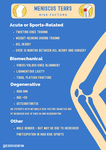
Meniscal knee injuries are commonly seen in MSK clinics and can impact athletic patients at all levels of sport or recreational activity. Injuries are seen...
Young athletes often complain of groin pain. When pain originates from the hip joint, clinicians refer to it as hip...
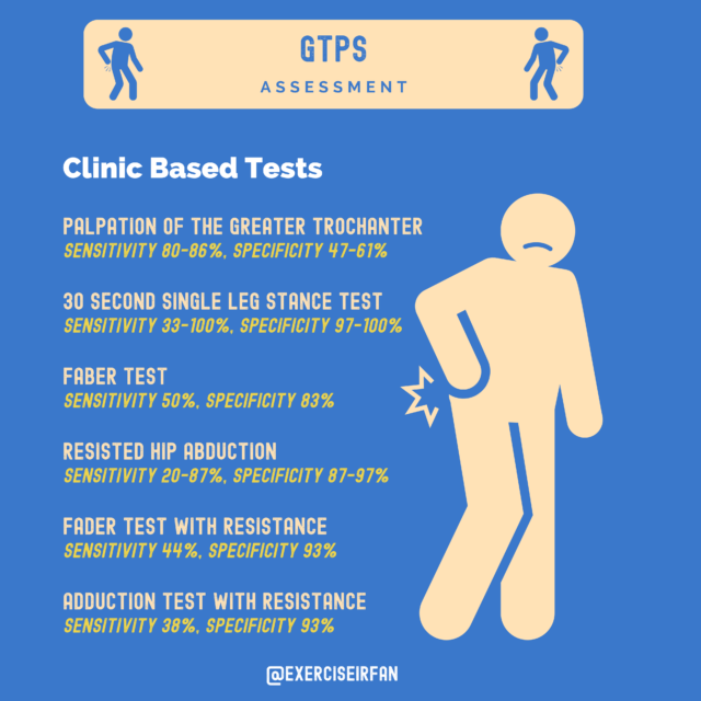
Greater Trochanteric Pain Syndrome (GTPS) is a common cause of lateral hip pain seen in the MSK clinic and can...
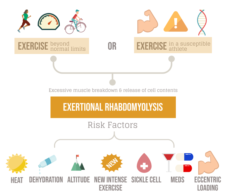
Exertional rhabdomyolysis (ER) is a condition that can impact athletes, patients and military personnel alike...
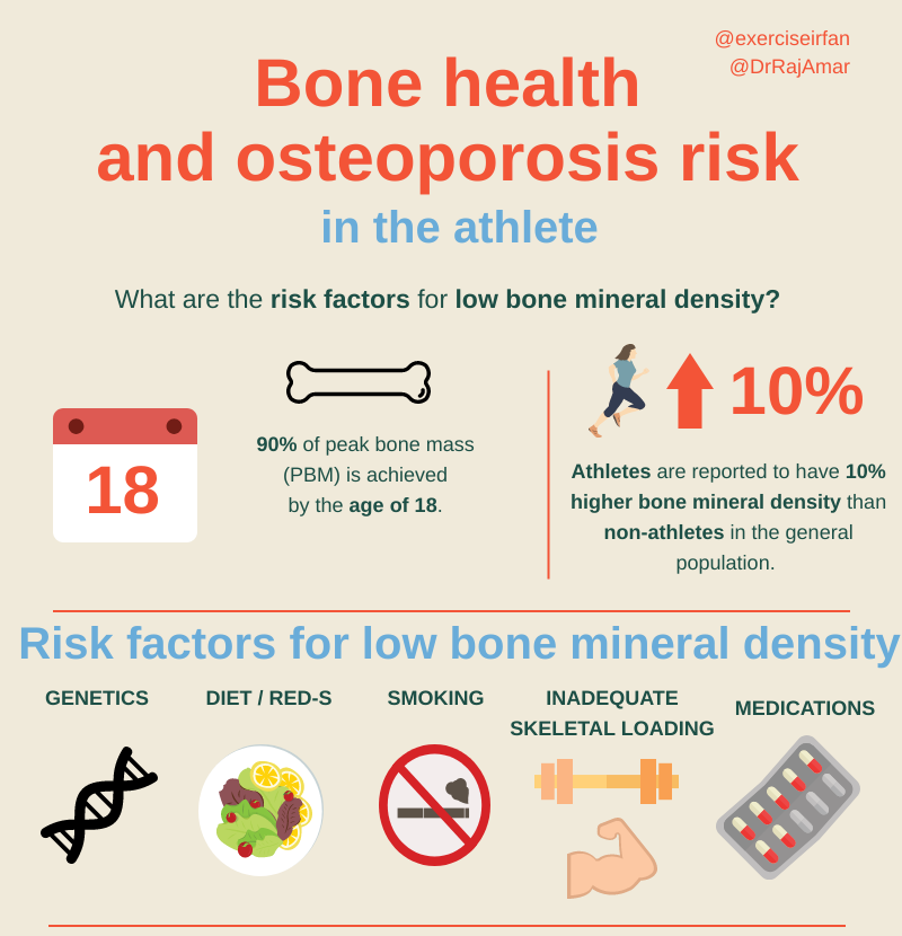
Bone health is a key area of development in the wellbeing of young athletes and is crucial for their safe training and...
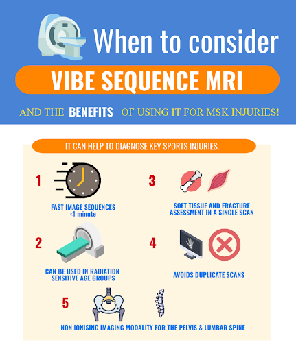
Volumetric interpolated breath-hold examination (VIBE) is an MRI sequence that is increasingly being used by...
Ankle sprains affect anyone and everyone along the activity spectrum, from semi-professional players and athletes to weekend warriors and...
For the athlete or exercising patient, injury, training load, genetics, and training type have traditionally been...
Anterior knee pain or “runner’s knee” is a common presenting complaint in the MSK clinic...
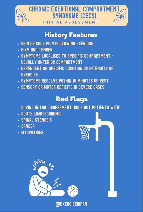
Exercise-induced leg pain (ELP) is a widespread condition among exercising adults that can hinder their ability to participate...
Module 6: Common knee conditions in the MSK clinic
6.1 The MSK playbook : Knee OA
6.2 – MRI of the knee – Live MRI tutorial to walk you through the key anatomy, how to identify common pathology and explain the scan to your patient!
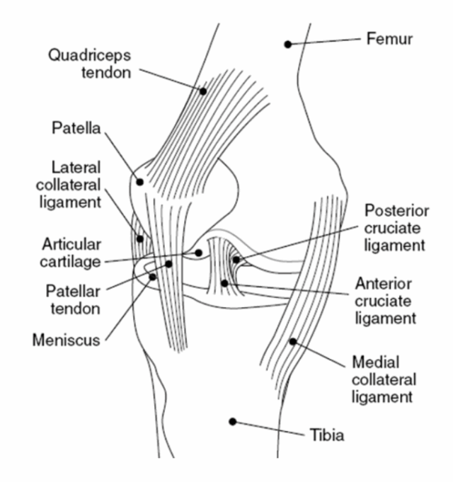
6.3 – MSK imaging series – (Fractures, dislocations, OA, meniscus injuries, ACL/PCL, MCL/LCL, osteochondral defects)
6.4 MRI of the knee – Learn the key views to be able to show your patient
6.5 The MSK playbook; Evidence updates on ACL injuries
6.6 The MSK playbook; Evidence updates on meniscus injuries
6.7 The MSK playbook; Evidence updates on the patello femoral joint
Module 7: Common Ankle conditions seen in the MSK clinic
7.1 – MRI of the Ankle – Live MRI tutorial to walk you through the key anatomy, how to identify common pathology and explain the scan to your patient!
7.2 – MSK imaging series – (Osteoarthritis, Ankle fractures, Achilles, Osteochondral injuries, tendinopathies, Sinus tarsi, Tarsal Tunnel syndrome)
7.3 - MRI of the Ankle – Learn the key views to be able to show your patient.
7.4 The MSK playbook; Lateral ankle injuries
7.5 The MSK playbook: Chondrocalcinosis
Module 8: Common foot conditions
8.1 – MRI of the Foot & Ankle – Live MRI tutorial to walk you through the key anatomy, how to identify common pathology and explain the scan to your patient!
8.2 – MSK imaging series - (Gout, OA, Stress fractures, Lisfranc/ midfoot injuries, sessmoiditis, metatarsalgia)
8.3 - MRI of the Foot & Ankle – Learn the key views to be able to show your patient.
8.4 The MSK playbook:Bone stress injuries in the athletic patient - bloods, DXA, imaging?
8.5 The MSK playbook: Exercise Induced leg pain
8.6 The MSK playbook: Plantar heel pain
Module 9: Common hip conditions in the MSK clinic
9.1 – MRI of the Hip – Live MRI tutorial to walk you through the key anatomy, how to identify common pathology and explain the scan to your patient!
9.2 – MSK imaging series - (Femoral acetabular Impingement, OA, Greater trochanteric pain syndrome, labral injuries, deep gluteal pain syndrome)
9.3 - MRI of the Hip – Learn the key views to be able to show your patient.
9.4 The MSK playbook; Greater trochanteric pain syndrome
9.5 The MSK playbook; Femoral acetabular impingement syndrome (FAI)
Module 10: The Medical work up
10.1 The MSK playbook : Exertional rhabdomyolysis
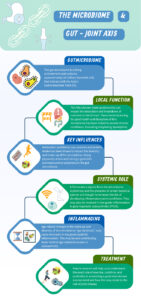
10.2 The MSK playbook: The Gut – Joint axis
*LIVE course content subject to change.
Comprehensive Sports Medicine Training
Courses Provided By Radiology Seminars
© 2026 Sports Imaging Institute. All rights reserved.